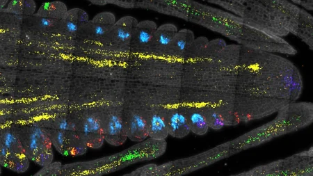
Oligodendrocytes are essential cells in the central nervous system that play a critical role in brain function. A new study featured in the Journal of Neuroscience reveals that mature oligodendrocytes have the remarkable ability to survive for much longer than previously thought. While younger cells typically die within 24 hours of a fatal trauma, mature oligodendrocytes were found to persist for up to 45 days. Understanding this extended lifespan may provide new insights into reversing the damage caused by aging and diseases like multiple sclerosis.
Oligodendrocytes are responsible for producing myelin, a protective membrane that coats the long connections between nerve cells. This myelin sheath helps in the efficient transmission of electrical signals along the axons, enabling effective communication between neurons. However, with age and neurodegenerative diseases, oligodendrocytes become damaged, leading to the breakdown of myelin production. This can result in the loss of motor function, feeling, and memory, as neurons struggle to communicate effectively.
Previous research suggested that damaged oligodendrocytes undergo a process called apoptosis, in which the cells self-destruct. However, the Dartmouth researchers found that mature oligodendrocytes may follow a different pathway before their eventual death. This unique mechanism raises questions about what changes occur in these cells as they mature, allowing them to persist longer than expected. Understanding this process could offer insights into potential interventions to either encourage or prevent this pathway in disease contexts.
The study’s lead author, Timothy Chapman, highlights the need for a nuanced approach to protecting oligodendrocytes. While efforts have typically focused on cultivating young cells and preserving mature ones, the findings suggest that aging cells may respond differently to treatments. Chapman emphasizes the importance of tailoring interventions based on the age of the cells, as young and old cells may require distinct strategies for preservation and regeneration.
The researchers developed a living-tissue model that allowed them to observe the death of a single oligodendrocyte and its surrounding cells’ response. They found that in a young brain, neighboring cells immediately replenished lost myelin when an oligodendrocyte died, whereas in an older brain equivalent, there was no regeneration. This model provided insights into the effects of aging on oligodendrocyte death and regeneration processes, shedding light on cellular-level mechanisms and potential controls.
Using innovative techniques such as a photon-based device called 2Phatal, the researchers induced fatal DNA damage in oligodendrocytes and monitored their response over time. The study revealed that mature cells did not resist damage but instead experienced an extended period of cell death that had not been observed previously. This prolonged death process raises questions about the functionality of myelin during this phase and whether accelerating the removal of dysfunctional myelin may be beneficial in certain contexts. Further research is needed to fully understand the implications of this extended cell death and its impact on oligodendrocyte function and brain health.


