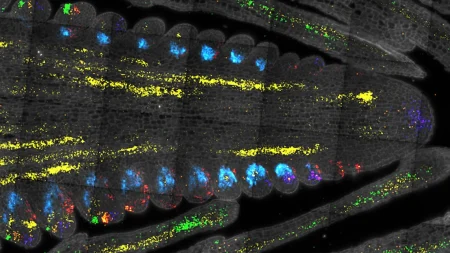
Understanding the behavior of molecules and cells within the human body is crucial for advancements in medicine. In a recent study published in Science Advances, researchers from Osaka University have introduced a method that provides high-resolution Raman microscopy images. Raman microscopy is valuable for imaging biological samples as it can offer chemical information about specific molecules involved in bodily processes. However, the Raman light emitted by biological samples is often weak, leading to poor image quality due to background noise.
The team of researchers has designed a microscope that can maintain the temperature of frozen samples during image acquisition, resulting in images that are up to eight times brighter compared to previous Raman microscopy images. By imaging frozen samples that are motionless, longer exposure times can be utilized without causing damage to the samples. This approach has led to higher signal-to-noise ratios, enhanced resolution, and larger fields of view. The technique does not require stains or chemicals to fix cells in position, providing a more representative view of cell behavior and processes.
The study also confirmed that the freezing process preserves the physicochemical states of various proteins, giving cryofixing an advantage over chemical fixing methods. Senior author Katsumasa Fujita highlights the complementary aspect of Raman microscopy to the imaging toolbox, as it not only offers cell images but also provides information on molecule distribution and chemical states. This detailed understanding is essential for researchers aiming to achieve a comprehensive view of biological processes.
The new technique can be combined with other microscopy methods for in-depth analysis of biological samples and is anticipated to have diverse applications in the biological sciences, including medicine and pharmaceutics. By providing high-resolution images and chemical information about molecules, Raman microscopy can contribute to a better understanding of cellular processes, which is essential for the development of new medical treatments and therapies. The ability to capture detailed images of molecular structures and behavior within cells can lead to significant advancements in various fields of science and medicine.
Overall, the development of high-resolution Raman microscopy images through the use of frozen samples represents a significant advance in imaging techniques for biological research. This novel approach enables researchers to obtain clearer, more detailed images of cellular processes and molecular interactions, facilitating a deeper understanding of biological systems. With the potential for widespread applications in medicine, pharmaceuticals, and other fields, this innovative method has the potential to drive further breakthroughs in scientific research and medical advancements.


