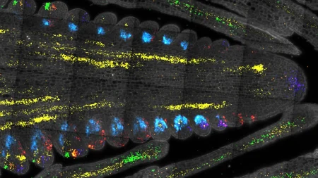
Researchers at MIT have developed a new technique that allows for nanoscale imaging of cells using conventional light microscopes, eliminating the need for expensive super-resolution microscopes. By expanding tissue before imaging it, researchers can achieve a 20-fold expansion in a single step, allowing for higher resolution imaging of organelles and protein clusters inside cells. This technique, known as expansion microscopy, was first developed in 2015 by Edward Boyden’s lab, and has since undergone modifications to achieve higher resolution images. With this new 20-fold expansion capability, researchers can visualize cell structures at a resolution of approximately 20 nanometers.
The expansion microscopy technique involves embedding tissue into an absorbent polymer, breaking apart the proteins that hold tissue together, and then adding water to swell the gel and pull biomolecules apart. The latest version of the technique uses a gel composed of N,N-dimethylacrylamide and sodium acrylate, which forms crosslinks spontaneously and exhibits strong mechanical properties. By removing oxygen from the polymer solution prior to gelation, the researchers were able to optimize the gel and polymerization process, resulting in a gel that can expand up to 20-fold without falling apart. This single-step expansion simplifies the imaging process and allows for high-resolution imaging with a conventional light microscope.
Using this technique, the researchers were able to image tiny structures within brain cells, cancer cells, and other cellular components. Synaptic nanocolumns, microtubules, mitochondria, and nuclear pore complexes were visualized with high resolution, providing insights into cellular organization and function. The researchers are also exploring the use of this technique to image carbohydrates known as glycans on cell surfaces, which could provide valuable information about cell-cell interactions and tumor cell organization. This simple and cost-effective method opens up new possibilities for studying nanoscale structures in cells across various biological contexts.
The democratization of imaging is a key aspect of this new technique, as it allows any biology lab to perform nanoscale imaging with existing equipment and standard chemicals. By providing a step-by-step protocol for the expansion microscopy process, the researchers aim to make this technique accessible to a wider range of researchers and labs. The use of off-the-shelf chemicals, common laboratory equipment, and standard microscopes enables labs to achieve high-resolution imaging that was previously only possible with specialized and costly microscopes. This advancement in imaging technology has the potential to revolutionize the study of nanoscale biology and expand our understanding of cellular structures and processes.
Funding for this research was provided by the U.S. National Institutes of Health, MIT Presidential Graduate Fellowship, U.S. National Science Foundation Graduate Research Fellowship grants, Open Philanthropy, Good Ventures, Howard Hughes Medical Institute, Lisa Yang, Ashar Aziz, and the European Research Council. The development of this new expansion microscopy technique represents a significant breakthrough in the field of biological imaging, offering researchers a simple and cost-effective way to visualize nanoscale structures within cells. By democratizing nanoscale imaging, researchers can access valuable insights into cellular organization, function, and pathology, paving the way for new discoveries and advancements in the field of biology.


