LMU researchers have developed a method to determine the reliability of labeling target proteins using super-resolution fluorescence microscopy, allowing for detailed observation of individual proteins within cells. The innovative RESI technique enhances the resolution of fluorescence microscopy to the Ångström scale by attaching DNA-conjugated marker molecules to specific proteins. A recent study published in Nature Methods details a technique that quantifies how effectively biomarker molecules bind to target proteins, enabling spatially resolved proteomics to understand protein presence, behavior, and changes in cellular environments. This assessment is crucial for accurate data analysis and comparisons between different binders and labeling conditions.
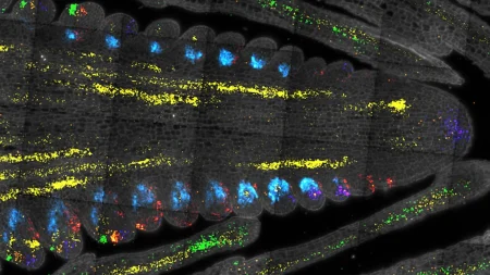
The method developed by Jungmann’s team involves the addition of a reference biomarker that emits a different color during microscopy, making successfully marked proteins appear in two colors. This dual-color approach was demonstrated using the membrane protein CD86, where the reference produced a pink fluorescence and the actual marker a bluish one, resulting in a pattern of pink and blue points of light. By calculating the ratio of double and single illuminated molecules, the marking efficiency can be determined, allowing researchers to assess how well the labeling has worked. This method offers several advantages, including the ability to work in vitro and in vivo, with various target molecules, biomarkers, and samples, and compatibility with different super-resolution techniques.
The new quantification method developed by Jungmann’s team has the potential to significantly expand the applications of super-resolution microscopy, particularly in specific biomedical contexts where the quantitative detection of proteins and processes is crucial. This includes cancer research, where understanding interactions between proteins on the cell surface and drugs at a molecular level is essential for developing new types of medication. The ability to accurately assess marker efficiency will ensure reliable data evaluation and enable precise comparisons between different research laboratories, ultimately enhancing the reliability and versatility of super-resolution microscopy techniques.
Overall, the study highlights the importance of assessing marker efficiency in super-resolution microscopy for quantitative and reliable protein analysis within cells. By developing a novel method to quantify binding efficiency, Jungmann’s team has opened the door to a wide range of potential applications in biomedical research, including cancer studies. This innovative technique offers a more versatile and reliable approach to assessing marker efficiency, allowing for more accurate data analysis and comparisons across different research settings. The future of super-resolution microscopy appears promising, with the potential for significant advancements in understanding protein interactions and cellular processes in various biomedical contexts.


