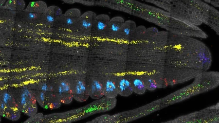
A team of chemists and bioengineers from Rice University and the University of Houston have made significant progress in creating a biomaterial for growing biological tissues outside the body. Their novel fabrication process creates aligned nanofiber hydrogels that could revolutionize tissue regeneration after injury and provide a platform for testing therapeutic drugs without using animals. Led by Professor Jeffrey Hartgerink, the team has developed peptide-based hydrogels that mimic the aligned structure of muscle and nerve tissues, which is crucial for their functionality.
For more than a decade, the team has been designing multidomain peptides (MDPs) that self-assemble into nanofibers resembling natural fibrous proteins in the body. In their latest study, they discovered a new method to create aligned MDP nanofiber “noodles” by dissolving the peptides in water and extruding them into a salty solution. This process creates aligned peptide nanofibers, like twisted strands of rope smaller than a cell. By increasing the salt concentration and repeating the process, they achieved enhanced alignment. The findings demonstrate that this method effectively guides cell growth in a desired direction, a crucial step towards creating functional biological tissues for regenerative medicine applications.
A key finding from the study was that excessive alignment of the peptide nanofibers hindered cell alignment, revealing that cells need to be able to “pull” on the nanofibers to recognize the alignment. When the nanofibers were too rigid, cells were unable to exert this force and organize themselves correctly. Understanding how cells interact with these materials at the nanoscale could have broader implications for tissue engineering and biomaterial design, leading to more effective strategies for building tissues. The research team included members from Rice University and the University of Houston, with support from various funding sources including the National Institutes of Health, the National Science Foundation, and the Welch Foundation.
The unexpected discovery during the study provides insight into cell behavior and could shape future tissue engineering and biomaterial design strategies. The ability to create aligned peptide nanofibers that guide cell growth in a specific direction is a significant advancement in regenerative medicine. The research team’s innovative approach to fabricating nanofiber hydrogels has the potential to revolutionize tissue regeneration after injury and offer a platform for testing therapeutic drugs. The study highlights the importance of achieving the right balance of alignment in peptide nanofibers to ensure optimal cell organization and functionality within biological tissues.
The collaborative effort between chemists and bioengineers to develop advanced peptide-based hydrogels is a promising step towards creating functional biological tissues for regenerative medicine applications. Identifying the optimal alignment of nanofiber hydrogels for guiding cell growth is a crucial aspect of tissue engineering and biomaterial design. This research has the potential to lead to more effective strategies for building tissues and could have broader implications for the field of regenerative medicine. The team’s groundbreaking findings offer new possibilities for tissue regeneration and drug testing, highlighting the importance of understanding cell behavior at the nanoscale in biomaterial design.


