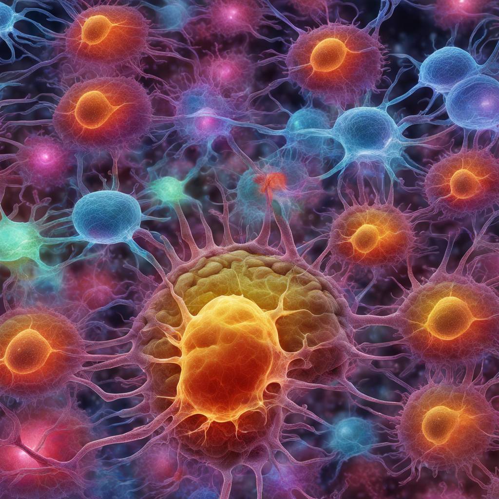
Researchers at the University of Wisconsin-Madison have developed a new tool that utilizes autofluorescence emitted by biological specimens to study the different states of stem cells in the nervous system. This innovative technique combines autofluorescence and genetic sequencing in single cells to examine the behavior of neural stem cells. While autofluorescence is often seen as a hindrance in research, the team found that it can be used to study stem cells’ dormant state, known as quiescence. The ability to identify and decode these autofluorescence signatures brightens the chances for studying how stem cells age and could lead to new insights into adult neurological diseases and aging.
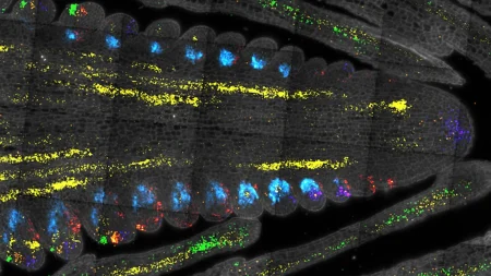
In a recent study published in Cell Stem Cell, the researchers focused on understanding the quiescent state of adult neural stem cells, which is crucial for the production of newborn neurons in the adult brain. They aimed to create a new tool that could identify if a cell is in a quiescent state or if it is activated and entering into the cell cycle. By analyzing the changes in light emission from specific proteins important to metabolism, the researchers were able to pinpoint the light signature associated with a target cell state. This approach allows for the non-destructive study of adult neural stem cells in various cell states, providing valuable insights into their behavior and function.
The development of this tool was a collaborative effort between Darcie Moore, a professor of neuroscience, and Melissa Skala, a biomedical engineering professor at UW-Madison. Skala’s lab specializes in fluorescence lifetime imaging to study autofluorescent signatures in single cells. By leveraging the changes in light emitted by different cellular components with quiescence, the team was able to identify unique autofluorescent signatures associated with specific cell states. This discovery not only provides a new tool for studying adult neural stem cells but also has the potential to expand beyond neuroscience to investigate other cell types, such as muscle stem cells.
Through RNA sequencing of mouse neural stem cells, the researchers confirmed the correlation between cell state and autofluorescence signatures. This validation further strengthens the utility of the new tool in studying cellular processes and identifying different cell states based on their functional measures rather than just the expression of individual proteins. By studying cells as they change over time without destroying them, researchers can gain a better understanding of dynamic systems and how functional measures evolve in different cell states. This approach has the potential to revolutionize the study of stem cells and improve our understanding of aging and neurological diseases.
The implications of this research extend beyond the field of neuroscience and have the potential to impact a wide range of scientific disciplines. Collaborations with other researchers, such as Colin Crist from McGill University, to investigate autofluorescence signatures in muscle stem cells highlight the broad applicability of the new tool. By uncovering the secrets of cellular processes through autofluorescence signatures, researchers can gain unique insights into cell behavior and function, paving the way for new discoveries in the study of stem cells and age-related diseases. This groundbreaking research was supported by grants from the National Institutes of Health, the Vallee Foundation, the Morgridge Institute for Research, and the Retina Research Foundation.


